We are still in the thick of winter here in South Carolina and the only green I still see outside comes from evergreens. One day on a walk, I looked up because a branch of a leyland cypress tree caught my attention. These branches are made up of flat needles that break down into fractals of smaller and smaller branches. I wondered if I could use the cast impression technique to view the stomata of the leyland cypress under my Foldscope 2.0! Read on to find out what I saw!
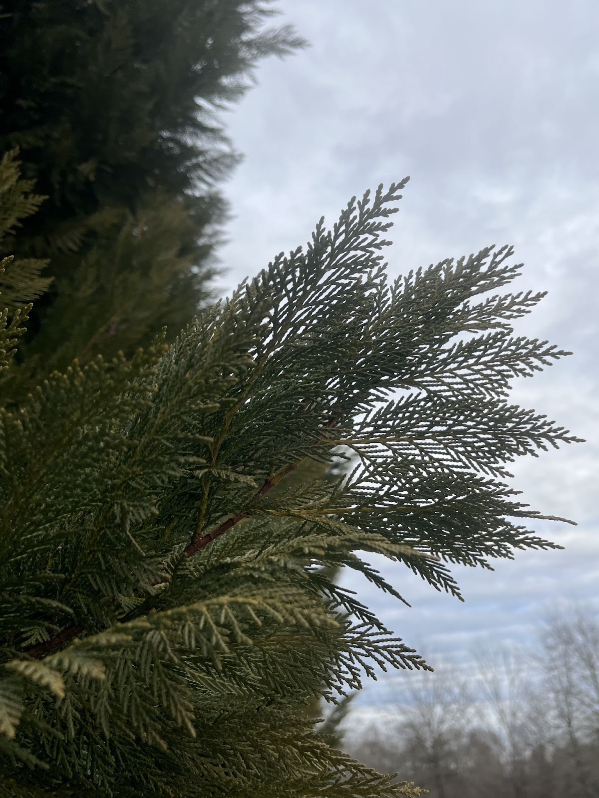
Figure 1. Picture of a branch of a leyland cypress tree
(Photo Credit: Holly A. Stuart)
Leyland Cypress Trees
Leyland cypress (scientific name: Cupressus x leylandii) are commonly used as a natural screen around houses for privacy because they are thick and fast growing trees. Their lush foliage, year round green color, and ability to grow even in poor soils makes them a popular landscaping choice.
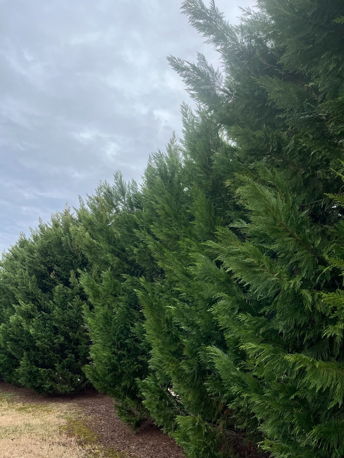
Figure 2. Picture of a row of leyland cypress trees
(Photo Credit: Holly A. Stuart)
Stomata
A stomata is a tiny pore, or opening, on the underside of a leaf where the plant’s gas exchange takes place. Guard cells around the stomata control when it is open or closed. What I love about stomata is that they look like miniature mouths!
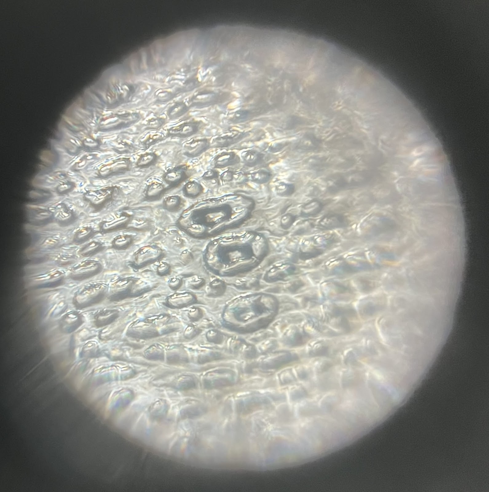
Figure 3. Picture of leyland cypress stomata viewed under a Foldscope 2.0 at 340X magnification
(Photo Credit: Holly A. Stuart)
Cast Impressions
So, how do you view stomata under a Foldscope when the leaves are too thick for light to pass through? You use the cast impression technique. This simple, yet effective, technique allows you to view surface features of opaque objects like thick leaves. Take a small piece of leyland cypress and paint a thin layer of clear nail polish on the underside of the leaf (the side where the stomata are more prevalent). When the polish dries, cover it with a piece of transparent tape and gently pull it off. Then, place the tape on a glass slide and it is ready for viewing.
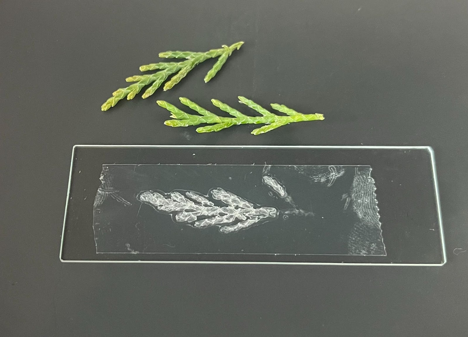
Figure 4. Picture of leyland cypress branches and glass slide with the nail polish and tape
(Photo Credit: Holly A. Stuart)
It is amazing just how clear the stomata is when viewed with a Foldscope 2.0 and an LED light module. I would say it rivals the image collected in this research paper that studied cypress tree stomata using a scanning electron microscope.
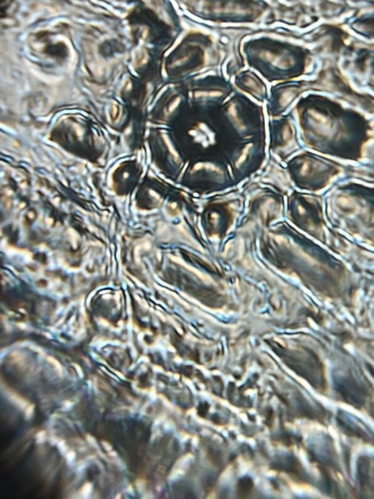
Figure 5. Picture of a leyland cypress stomata viewed under a Foldscope 2.0 at 340X magnification plus 5X zoom on my phone
(Photo Credit: Holly A. Stuart)
One thing I am now curious about is the irregularity of the stomata placement on the leaf. Some are clustered together, while others are singular and scattered. It makes me wonder if this is a result of the position of the branch on the tree, how much sunlight the branch receives, or some other environmental factor.
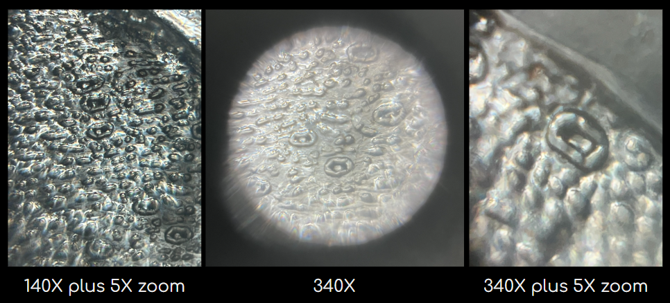
Figure 6. Picture of leyland cypress stomata viewed under a Foldscope 2.0
(Photo Credit: Holly A. Stuart)
Have you looked at stomata under a Foldscope 2.0? Use your Foldscope to dive into the microscopic world and find the beauty that is there waiting for you. Share your microscopic images and thoughts on the Microcosmos. Be sure to tag us on social media when you post the results of your explorations, creations, and discoveries! We love to see how Foldscopers around the world are using their Foldscopes in new and innovative ways!
Facebook: @Foldscope
Instagram: @teamfoldscope
Blue Sky: @teamfoldscope.bsky.social
TikTok: @foldscope
Threads: @teamfoldscope
Twitter: @TeamFoldscope
Sources:
https://kids.kiddle.co/Leyland_cypress
https://kids.kiddle.co/Stomata
https://link.springer.com/article/10.1007/s00468-023-02424-2


