Question:
Do artificial flowers look like real flowers under a microscope?

Hands-on experiences are the best way to learn about the scientific world. Dr. Marie Clark Taylor understood this and promoted teaching botany by using real botanical samples and light microscopes in classrooms.
It is important for students to interact with real specimens to learn:
- about variations in plant structures
- how to handle living specimens
- how to do dissections
- how to prepare slides for microscopy studies.
This activity is an opportunity for students to put Dr. Marie Clark Taylor’s teaching methods into practice! Can you and your students use a Foldscope to tell the difference between real plants and artificial plants?
Background:
Dr. Marie Clark Taylor (1911-1990) was a botanist who studied the effect of light on plant growth. But she also had an impact on science education! Dr. Taylor felt that it was important for high school teachers to use real botanical samples and light microscopes in their classrooms. So, the fact that we are going to be using Foldscope microscopes with this activity is thanks to Dr. Taylor!
In 1941 Marie Clark Taylor became the first African American woman to earn a PhD in science from Fordham University in New York. She was actually the first woman of any race to reach this level of academic achievement from Fordham University! Think about what that must have felt like - this was before the Supreme Court case of Brown vs. the Board of Education, the Civil Rights Act, and before some colleges and universities were even admitting black students into their schools.
Before and after earning her doctorate, Dr. Taylor taught high school science classes. The experience led her to develop some innovative teaching methods. She knew that learning botany by looking at pictures from textbooks wasn’t good enough. By bringing real plant specimens and light microscopes into her classroom, her students gained the benefit of being able to observe cellular structures and processes in real time.
This was considered a novel approach to teaching. And during the 1960s, it caught the attention of President Lyndon B. Johnson. He requested that Taylor teach educators all across the United States about the importance of using living plants and light microscopes in their classrooms! With the support of President Johnson, Dr. Taylor led programs that taught other teachers how to use these techniques which helped spread this style of teaching all around the world.
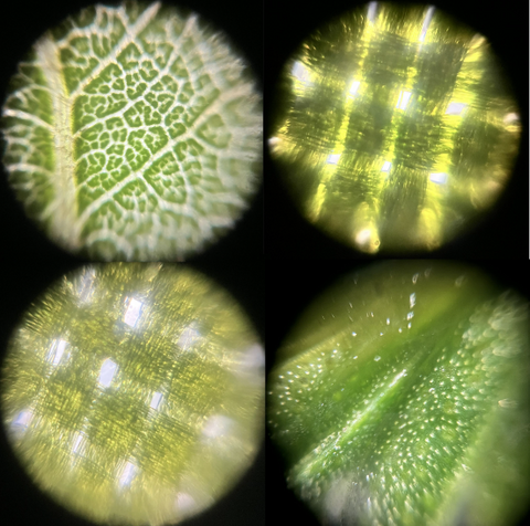
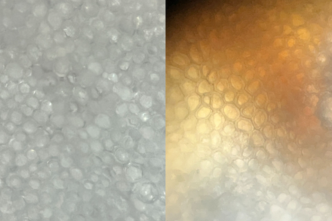
And now, you get the benefit of using microscopes and real plants in your classroom - which is a fun and interesting way to learn science! This activity will use a Foldscope (which is a type of light microscope) to compare real plants and artificial plants. While they may look alike on the surface, you will quickly see that they are very different on the microscopic level!
Materials/Procedure:
This activity uses living plants, so be sure to check for any potential allergies. If you do have students with allergies, they can be responsible for creating slides of the artificial plant specimens. To create the drawings of the real plant structures, they can base their observations off of images captured with a phone or tablet.
- Materials:
- Science notebook
- Pen/pencil
- Colored Pencils
- Foldscopes
- LED Light Modules
- Lens Kit (optional)
- Slides: glass,paper, or blank trading cards
- Cover slips: glass or clear stickers
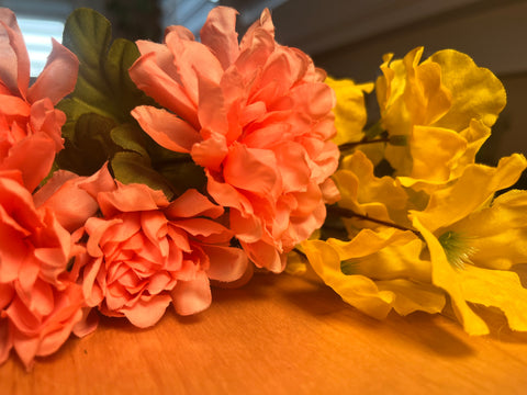
- A variety of artificial plants
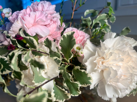
- A variety of real plants
- Dissecting Kit
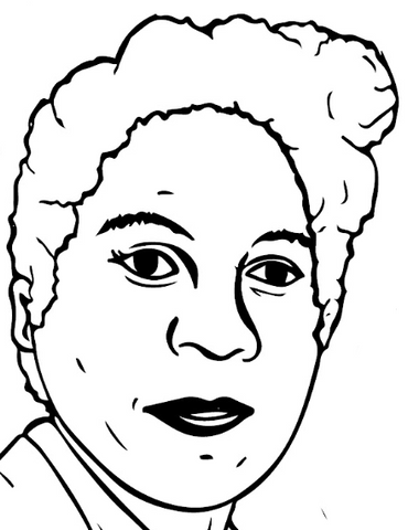
- Dr. Marie Clark Taylor Coloring Sheet
- Explain to the students that they are going to be comparing real and artificial plant parts under a Foldscope.
- Dry Mount Slides with real flowers:
- Place a flower petal, thin leaf, stamen, thin stem cross section, or any other plant parts that you choose, on a slide
- Use the dissecting kit to make sure that the samples you use are very thin.
- The scissors can cut small pieces off of the plant.
- The scalpel can cut thin slices of the stem.
- The tweezers can pick up the tiny pieces and place them on the slide.
- Cover the specimen with a glass coverslip or clear sticker
- Repeat with artificial flowers.
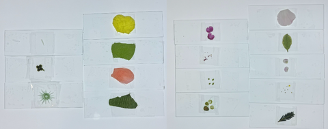
- Place the slides in the Foldscope and draw what you see in your science notebook. Use colored pencils so that you can capture the colors you see from the microscopic view.
- Label your illustrations. Did you see cells? Vascular bundles? Pollen? Or something else? Write it in your science notebook.
- Do you see any patterns between your microscopic observations? Create Venn diagrams to show similarities and differences between the real and artificial plants.
- Read over and color in the Color Me PhD coloring sheet about Dr. Marie Clark Taylor.
- Do you think that you could learn about the structures of plants if you didn’t have microscopes? Why or why not?
This activity was created to bring attention to the amazing work of Dr. Marie Clark Taylor for Black History Month. Use these ideas to inspire new ways of using microscopes in your classes every day! This way, you can celebrate the legacy of Dr. Taylor all year long, not just in February!
Another scientist (and artist) who celebrates the work of scientists of color is Dr. Julie Rorrer. She is the creator of the website Color Me PhD. On this site you can find coloring pages that provide information about scientists, the work that they did, and their lasting contribution to science. Dr. Rorrer feels that creating these coloring pages is important because she wants to “spark interest in science and engineering for all young students, regardless of gender, race, or socioeconomic status.” Her inspiration for creating Color Me PhD came about when she decided to create illustrations that explained her research to children she mentored. Do you think that you could create a coloring page that shows some of the ways you can use a Foldscope?

Extension:
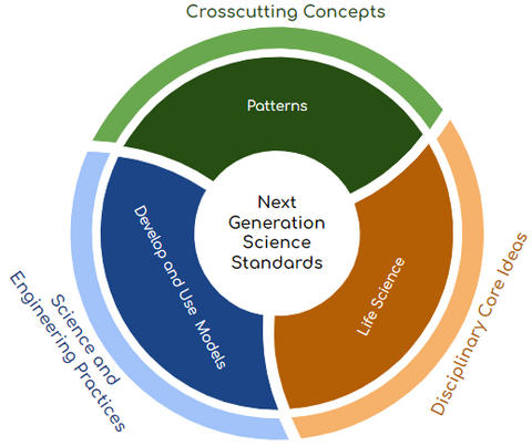
This blog ties together the three dimensional framework of the NGSS. It covers the Disciplinary Core Idea of Life Science. Students will see the Crosscutting Concept of Patterns. This activity is also a way for students to deepen their understanding of the Science and Engineering Practice of Develop and Use Models.
However, this exploratory activity can go beyond the science classroom. Join forces with:
- a Social Studies teacher to research the time period that Dr. Taylor lived in to better understand her struggles,
- a Math teacher to use the tools of math (rulers/compasses/etc.) to improve upon the diagrams and drawings in the science notebook, ensuring that they are drawn to scale,
- an ELA teacher to write an argumentative essay on the importance of hands on experiences and science equipment in the classroom,
- and a Visual and Media Arts teacher to create coloring pages inspired by the style of Color Me PhD!
Connect:
Could you tell which Foldscope images were from real plants and which ones were from artificial plants? How did your samples compare to the ones in this blog? Share your observations, discoveries, pictures, and interdisciplinary extension activities with the Foldscope community. Submitting your Foldscope images related to the topic to the Microcosmos will help build up a strong scientific database that can help support new and innovative scientific research!
Sources:
https://www.danforthcenter.org/news/celebrating-black-plant-scientists-throughout-history/
https://www.slbi.org.uk/assets/uploads/2020/10/Black-Botanists-Powerpoint-Presentation-Oct-2020.pdf
https://dbg.org/marie-clark-taylor/
https://www.wowstem.org/post/marie-clark-taylor
Dr. Marie Clark Taylor Coloring Sheet: https://drive.google.com/file/d/1tVR5y8lGNrTRnyUSmXgEtoT9Qkyc2lHx/view


