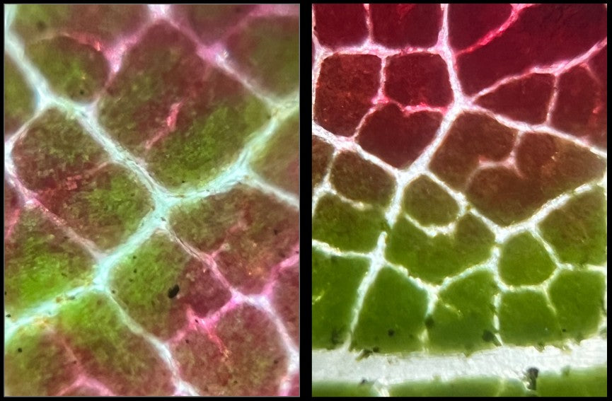
You’ve probably heard the saying, “An apple a day keeps the doctor away.” It turns out that in addition to being healthy, apples also have huge economic benefits. In 2020, apples generated $78 billion worldwide! So, it was with some new found respect that I decided to look at an apple peel under my Foldscope.
What is the apple peel made of?
The peel of an apple is called the exocarp. The exocarp is a multilayered structure that includes the cuticle, epidermis, and hypodermal tissue. The exocarp plays a very important role in protecting the fruit. Due to the economic importance of the apple market, researchers spend a lot of time studying the structural integrity of the exocarp. This is because the more protection the exocarp can provide the fruit, the better the apples will look when they reach the market.

What Did I See?
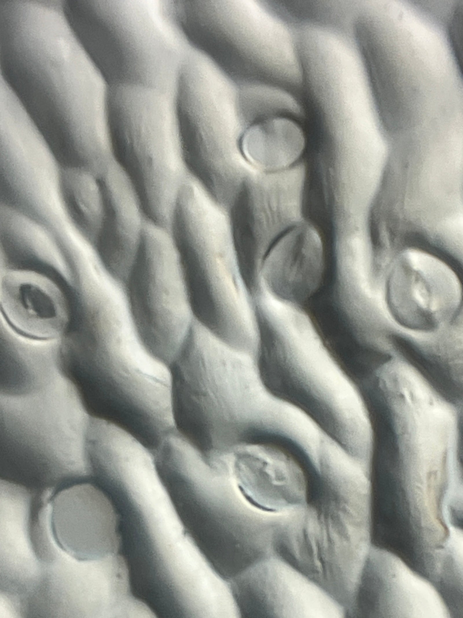
To prepare the slides, I took a very thin, translucent layer of a Granny Smith apple peel, cut it into a small strip, and placed it on a glass slide (of course you can always use paper slides, too!). To hold the apple skin sample in place on the slide, I covered it with a clear sticker.
The individual plant cells were visible at 140X magnification, but the finer details became more evident at 140X magnification plus 5X zoom on my phone. With the extra magnification from my phone, it looked like some cell walls were thicker than the others.

What Did I See With Staining?
To prepare these slides, I took a very thin, translucent layer of a Granny Smith apple peel, cut it into a small strip, and placed it on a glass slide (remember, you can also use paper slides). Then I added a drop of diluted methylene blue vital stain. To seal the apple skin sample in place, I covered it with a clear sticker.
Again, the cell walls were visible at 140X magnification, and seen with more detail at 140X magnification plus 5X zoom on my phone. The methylene blue vital stain enhanced the cell walls in the pictures which made them stand out against the interiors of the plant cells.

The microscopic view of the apple peel reminded me of the skin on the back of my hand viewed under a magnifying glass. Isn’t it interesting how patterns in nature appear in so many different locations, ecosystems, and organisms? What does the microscopic view of an apple peel make you think of? Take a look under a Foldscope and share your microscopic images and thoughts on the Microcosmos. Be sure to tag us on social media when you post the results of your explorations, creations, and discoveries! We love to see how Foldscopers around the world are using their Foldscopes in new and innovative ways!
Facebook: @Foldscope
Twitter: @TeamFoldscope
Instagram: @teamfoldscope
Sources:
https://fruitgrowersnews.com/news/global-apple-market-reached-78m-set-to-continue-moderate-growth/


