Question:
How do plants defy gravity?
Vascular plants have the ability to pull water from the ground and up to the leafy canopy against the force of gravity. For example, a 70 foot tall red oak tree absorbs about 100 gallons of water per day and pulls it up at a rate of 92 ft/hr!
Let's learn more about the structures inside of vascular plants that can move water upward without the use of a pump!

Background:
An important classification used for plants is whether or not they have a vascular system. The plant vascular system includes xylem and phloem, structures that move water and nutrients throughout the plant.
Xylem are tubelike structures that move water up through the plant from the roots to the leaves. Xylem is made of dead cells that are responsible for transpiration (the evaporation of water through the leaf stomata). There are no cell walls inside of the xylem, making it a hollow tube (like a straw) that also provides structural support to the stem of the plant.

Phloem are the tubelike structures that move sugars up or down the plant to wherever they are needed. Phloem is made of living cells, has sieve-like cell walls, and is responsible for translocation (moving food around).

Xylem is able to raise water up so effectively because of evaporation and hydrogen bonding. The water that reaches the leaves evaporates through the stomata. The evaporating molecules of water are attracted to the water behind them that are still in the plant through hydrogen bonding. This attraction is very strong. As the water molecules evaporate away, they tug on the molecules behind them. These water molecules get pulled through the stomata, too, and the process continues over and over again! The combination of evaporation and hydrogen bonding provides enough power to steadily pull the water up through the xylem tubes!
Phloem is able to get sugar to the parts of the plant that require it through the process of osmosis. When a leaf has made sugar through photosynthesis, the high concentration of sugar in the leaf gets transferred to the more dilute phloem cells. Water from the xylem flows in to dilute the concentrated sugar in the phloem, resulting in an area of high pressure. The high pressure in the phloem is released by moving the sugars and water to a location in the phloem next to a cell that needs food. The sugar goes to the cell, and the excess water flows back to the xylem.
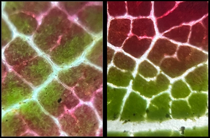
The xylem and phloem tubes are found in bundles within the stem of a plant. A microscopic view of a cross section of the stem reveals their presence as a group of circles tightly packed together. This perspective shows the “tops” of the tubes. The addition of a methylene blue stain to the sample makes the xylem and phloem stand out and much easier to identify.

You can determine if you have a monocotyledon (a flowering plant with an embryo that bears a single seed leaf) or dicotyledon (a flowering plant with an embryo that bears two seed leaves) plant by the arrangement of the vascular bundles. Monocotyledons have the vascular bundles randomly spaced within the stem. Dicotyledons have the vascular bundles arranged in a circle along the outer edge of the stem.
This activity was designed with springtime in mind and uses asparagus plants because they are plentiful in the spring. When studying them under the Foldscope, can you determine if they are monocotyledons or dicotyledons?
Directions/Procedure:
Here is what you will need and what you will do in order to complete the activity with your students..
Materials:
- Science notebook
- Pen
- Microtome and/or sharp knife for making thin slices of your sample
- Asparagus stems (or other seasonally available vascular plant stem)
- Methylene blue vital stain
- Disposable pipettes
- Water
- Slides
- You can use glass or paper slides
- Slide covers
- You can also use clear stickers or clear tape
- Microscope (or a Foldscope)
- Place the asparagus stem in the microtome and prepare thin cross sections of the stem.

- Place the asparagus stem in the microtome and prepare thin longitudinal sections of the stem.
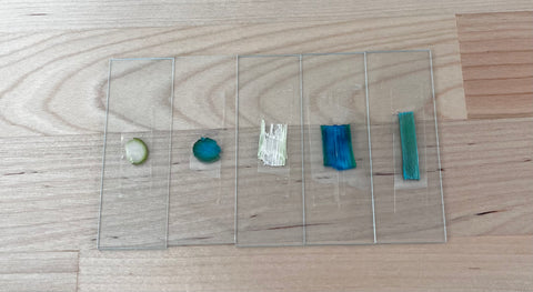
- Prepare slides of both the cross section and the longitudinal section of the asparagus stem.
- Take a cross section of the asparagus stem and stain it with the methylene blue vital stain.

- Place a drop of methylene blue on the cross section
- Immediately rinse the stem with water using the pipette.
- When the water runs clear from the rinse, prepare a slide of the stained cross section of stem.
- Repeat the staining with the longitudinal section of the asparagus stem.
- View all of the slides under the Foldscope.

- Answer the following questions in your science notebook:
- Can you see the vascular bundles?
- How did the cross section view differ from the longitudinal view?
- Did the staining help you to see the xylem and phloem more clearly?
- Using your science notebook and pen, draw and label what you saw in your Foldscope. (If you are in a dark room, it might help to try projecting the image onto your paper and tracing it!)
Do you think that the vascular bundles of other plants differ from those of the asparagus plant? How and why?
Do you think that the vascular bundles of the asparagus change throughout its life cycle? How? Can you design an experiment to test your hypothesis?
Real World Topic Scientists & Artists:
Katherine Esau (1898 - 1997) was an author, researcher, and professor at both UC Davis and UC Santa Barbara. Her areas of interest included plant viruses, developmental and pathological plant anatomy, and ultrastructure of phloem. As a botanist, Esau’s research focused on phloem and its relationship to plant disease. She discovered that the curly top virus affecting sugar beets spread inside the plant through the phloem. How can your observations of phloem contribute to Esau’s work?
Regina Marzlin is a Fiber Artist from Nova Scotia, Canada. Marzlin began working with textiles in 2004 because she enjoyed using fabrics and stitching to create different textures. Marzlin currently has a quilt called “Xylem and Phloem” featured in the Studio Art Quilt Associates Global Exhibition: Microscapes. Marzlin is quoted as saying “Inspiration is all around me and in me.” What does the microscopic inner structure of plants inspire you to create?
Extension:
This blog ties together the three dimensional framework of the NGSS. It covers the Disciplinary Core Idea of Life Science. Students will see the Crosscutting Concept of Structure and Function. This activity is also a way for students to deepen their understanding of the Science and Engineering Practice of Engage In Argument From Evidence.

However, this exploratory activity can go beyond the science classroom. Join forces with:
- a Social Studies teacher to investigate the historic impact of the sugar trade on global society (the sap that creates sugar comes from the vascular system of plants),
- a Math teacher to determine the density of vascular bundles in different plants,
- an ELA teacher to create Haibun poems encouraging students to make visual connections to being inside of the xylem or phloem,
- a Design teacher to create functional models of vascular bundles using materials appropriate to the task.

Connect:
Make sure to share your observations, hypothesis, results, and interdisciplinary extension activities. Submitting your Foldscope images of vascular bundles, xylem, and phloem to the Microcosmos will help build up a strong scientific database that can help support new and innovative scientific research!
Sources:


