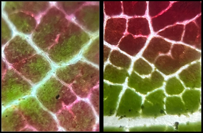
Recently I came upon some small flowers growing in the grass. There were groups of them and while each had a bright yellow center, the outer edges of the petals were white, light purple, or fuchsia. Of course I had to put them under my Foldscope 2.0. Read on to learn how to prepare flower petals for microscopic observation and to see what I saw!

Preparing flower petals for microscopy is a simple procedure that yields big results! If the flower petals are thin enough to let light pass through them, you can use a slide prep technique called a dry mount. This is just another way of saying “put the dry sample directly onto the slide.”
This is how I prepare my flower petal slides. I cut a small piece of flower petal off of the flower with a pair of scissors. Then I secure the flower petal piece onto a glass slide using a cover slip or clear sticker.

I chose a piece of the flower petal that included the area where the yellow color in the center transitioned into the color on the outside of the flower.

I chose this part of the flower petal because I wanted to see what the color transition looked like on a microscopic scale. Even though the macroscopic view of the flower petal looks like it is a clear line between the two colors, the Foldscope reveals a different view. It is actually a subtle transition from one color to the next.

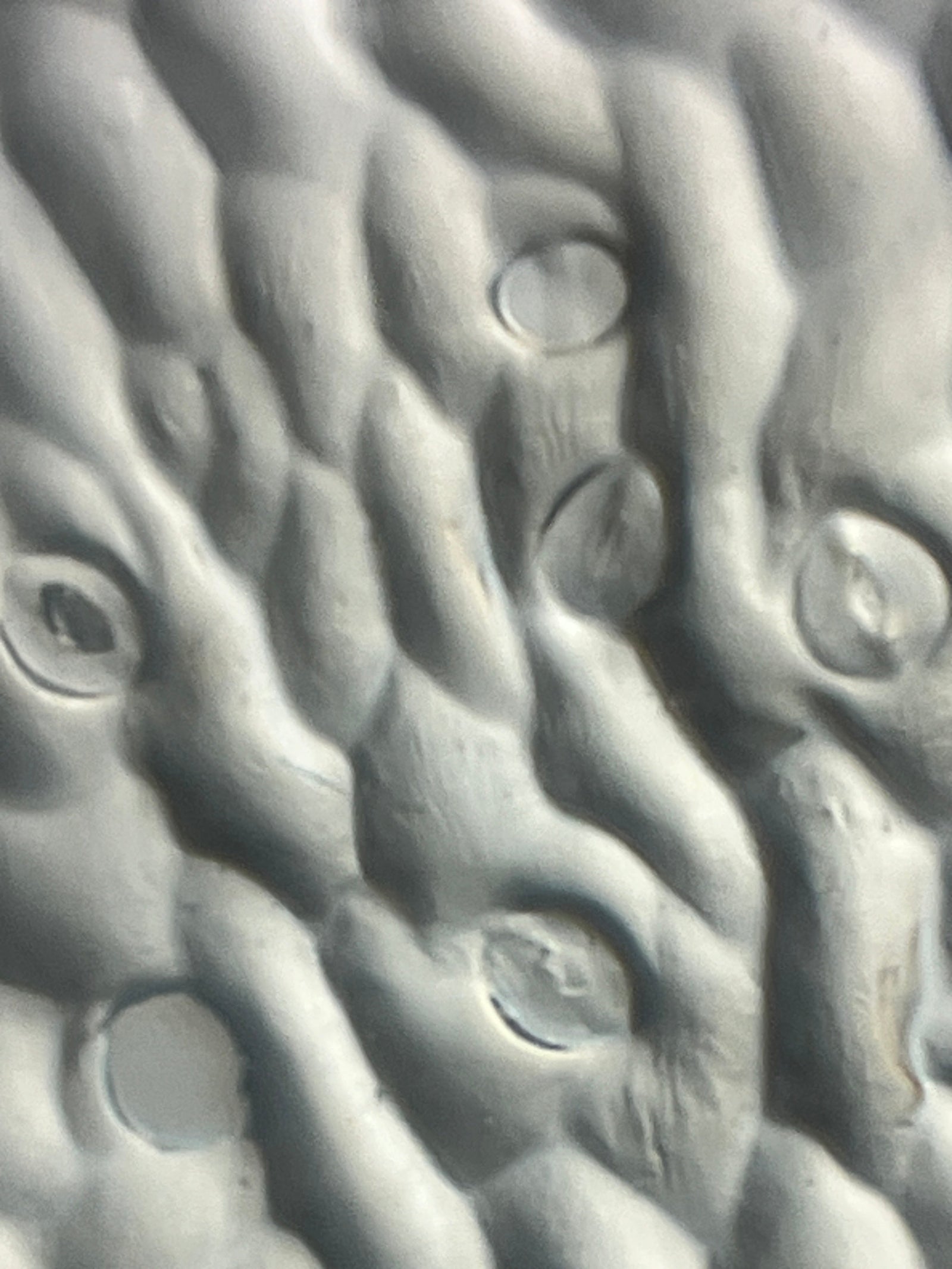
When preparing slides don’t worry if the flower petal doesn’t lie completely flat on the slide. These imperfections can actually provide some visual interest as you look through the Foldscope. Even the smallest of folds or wrinkles in the flower petal adds a three dimensional quality to the microscopic image. The thin and delicate flower petal can look like a thick folded blanket.

When looking at flower petals under a Foldscope, don’t forget the edges. You can discover some surprising features and get interesting light effects at the edge of the petal.

I hope this blog post inspires you to try making dry mount slides of flower petals too! Use your Foldscope 2.0 to dive into the microscopic world and find the beauty that is there waiting for you. Share your microscopic images and thoughts on the Microcosmos. Be sure to tag us on social media when you post the results of your explorations, creations, and discoveries! We love to see how Foldscopers around the world are using their Foldscopes in new and innovative ways!
Facebook: @Foldscope
Twitter: @TeamFoldscope
Instagram: @teamfoldscope
Threads: @teamfoldscope


